
Laparoscopic Gastric Bypass Steps

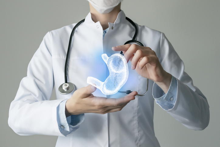
Laparoscopic gastric bypass surgery, otherwise known as GBP, is a surgical procedure that involves constructing a gastric pouch, which results in remarkable and long-lasting weight loss for severely obese patients. The surgical procedure results in a new stomach pouch that is affixed to a piece of the small intestine, which is connected directly onto the new stomach pouch. After this surgery, patients are able to only hold approximately 1 to 2 ounces of food when eating, as the stomach is now reduced to a significantly smaller size. The new stomach pouch can range from the size of a bowling ball or can be as small as the size of a golf ball, depending on the severity of the case. Laparoscopic gastric bypass surgery is a significant surgery, given the fact that the stomach generally can hold approximately 48 ounces of food otherwise. Therefore, this type of surgery is typically ideal for severely obese patients.
After the surgery and the patient consumes their food, the food enters the small stomach pouch and then goes directly into the center section of the small intestine. As a result, smaller amounts of food is consumed and fewer calories are absorbed, which then leads to weight loss. The surgery essentially produces two channels within the intestinal tract: (1) for food and (2) for digestive juices.
Steps of Laparoscopic Gastric Bypass Surgery
First, tiny incisions are created along the abdominal wall in order to reach the abdominal cavity. Trocars, which are surgical tools that are access devices allow the surgeon to pass through the inciscions seamlessly to effectively get to the abdominal cavity. Once the abdominal cavity is accessed, the abdomen is then expanded, similarly to a balloon, with carbon dioxide (CO2) gas, which results in the abdomen to swell and raises the stomach wall away from the small intestine, along with the other organs. This will allow your surgeon to get a better view of the abdominal cavity and organs, while using a laparoscope. A laparoscope is a thin, long tool that has a tiny video camera and light attached to the end.
Next, the procedure involves utilizing the omentum, which is the large fold of fatty tissue that acts as a cover and protects the intestines and organs that are located in the lower abdominal area, along with the transverse colon, which is part of the large intestine that is located over the paper part of the abdominal cavity. These organs and tissues are then lifted up towards the head in order to examine and open the ligament of Treitz. The Treitz is a thin band of tissue that links the duodenum to the diaphragm. This allows the surgeon to lift the transverse mesocolon, which is the fold that links the transverse colon and the abdominal cavity, and then creates an opening. The opening allows the bottom of the stomach to be identified.
The next part of the procedure requires the jejunum to be divided, which is the center segment of the small intestine, into two separate sections. The two sections, which are the biliopancreatic limb and the Roux-limb eventually become the alimentary limb, which will be connected to the new stomach pouch. As the intestines are now separated, your surgeon will then measure the jejunum along the digestive tract. This will allow the surgeon to determine the length of your alimentary limb. The smaller part of the jejunum is closed and fastened to the longer part, which is what was measured out.
Next, the surgeon creates an anastomosis. An anastomosis involves connecting two elements in the body, which in this situation, is putting two pieces of the jejunum together. In this case, this is referred to as a jejunojejunostomy. Once the small intestine is connected to one another (generally by suturing and stapling), the jejunojejunostomy is then secured for the prevention of an internal hernia. The small intestine is then positioned in the back of the colon, which will eventually be connected to the stomach pouch. Once this step is done, the stomach will then be split about 1-2 inches underneath the area of the esophagus connected to the stomach by creating an opening in the open space of the gastrohepatic ligament. The gastrohepatic ligament is what connects the stomach and the liver. As the minor curvature of the stomach is accessed, there will be a space located in the back of the stomach that gives access to the smaller pouch. The stomach is then divided further with staples in order to achieve a completely separate section. The smaller section located above the stomach that is still connected to the esophagus is essentially your newly created stomach, otherwise referred to as the gastric pouch. The gastric pouch is able to hold about 1 to 2 ounces of food.
After the gastric pouch is created, the rest of the jejunum is positioned behind the colon and the older portion of the stomach so the gastric pouch can be efficiently attached. Once this step has been completed, the new attachments are then checked to ensure security and a leak check is usually performed. The rest of the incisions will then be secured with sutures and your anesthesia will stop being administered so you are able to wake up once your surgery has been completed.
Contact Us to Schedule a Consultation
Interested in gastric bypass surgery and the steps involved? Our team is here to help give you as much clarity as you need regarding the gastric bypass steps and whether you are an ideal candidate for the surgery. Contact our team at Weight Loss Los Angeles to learn more!

Discover the Best Los Angeles Weight Loss Clinic – A New Approach to Medical Weight Loss
Body and Mind: This is the True Meaning of Weight Loss
Why Zepbound Could Be the New Ozempic

What Is Mounjaro? Exploring a Revolutionary Weight Loss Treatment for Type 2 Diabetes

How much weight can I lose with Wegovy?
How Does Wegovy Work?
How Does Saxenda Work?

Everything you need to know about Mounjaro Pen for weight loss
Everything you need to know about Saxenda Weight Loss